


The pancreas is a very important organ in our body, with a dual role. It is located in the posterior part of the abdomen (retroperitoneum), behind the stomach and intestines, and above the large arterial and venous blood vessels. Due to the organ’s location, treating pancreatic diseases and performing necessary surgeries on it requires a high level of expertise and precision.
In this article, we will discuss the symptoms, diagnosis, and treatment of the most well-known pancreatic diseases: pancreatitis and pancreatic cancer.
What does the pancreas look like?
The pancreas has a glandular structure with two parts: exocrine and endocrine. The exocrine part of the pancreas produces enzymes essential for digesting the food we consume and is connected through the main pancreatic duct to the intestines, where pancreatic enzymes reach their mature form and break down proteins and fats from food into nutrients, which are then absorbed in our intestines.
The endocrine part of the pancreas consists of numerous islets where specialized cells are grouped. These cells produce the main hormones for regulating blood sugar levels, insulin, and glucagon.
What is pancreatitis?
Inflammation of the pancreas is called pancreatitis, and acute and chronic forms are distinguished.
Acute pancreatitis
Acute pancreatitis occurs when pancreatic enzymes are activated within the organ itself, leading to damage to the glandular structures. This condition has various causes:
- Biliary pancreatitis occurs when a stone in the bile duct prevents the passage of pancreatic juices into the intestines, causing the enzymes to activate within the organ;
- Ethylic pancreatitis occurs due to the intake of a large amount of alcohol;
- Metabolic pancreatitis occurs due to high levels of fat in the blood;
- Drug-induced pancreatitis occurs when the intake of specific medications leads to enzyme activation and pancreatic damage;
- Idiopathic pancreatitis involves an unknown cause of the disease’s onset.
In most cases, acute pancreatitis is treated with medication therapy along with restricted food intake, while complications of acute pancreatitis (necrosis, formation of collections) can be treated surgically.
Chronic pancreatitis
Chronic pancreatitis is a prolonged form of the disease that arises from repeated acute episodes, most commonly occurring in patients with long-term excessive alcohol consumption.
Symptoms of this disease often include:
- Weight loss;
- Diarrhea accompanied by fat loss;
- Pain, which is most often the reason for surgical treatment.
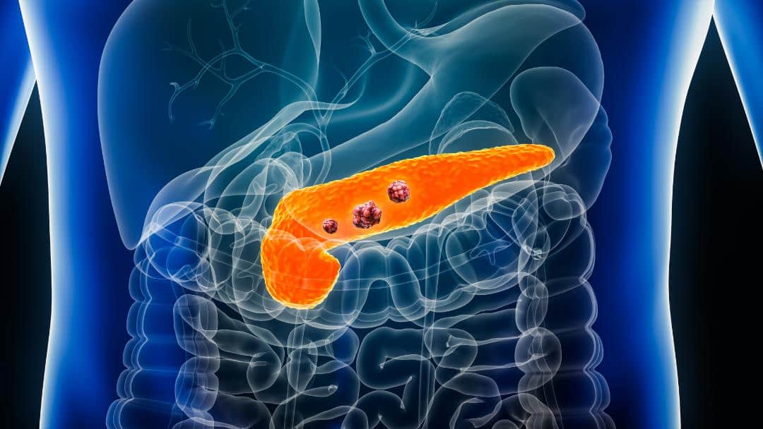
How does pancreatic cancer manifest?
Pancreatic cancer (adenocarcinoma) is a malignant tumor of the exocrine part of the pancreas. The frequency of this tumor is increasing in Western Europe, primarily due to more prevalent risk factors and more effective diagnostics of this disease.
Risk factors for pancreatic cancer
their sixth and seventh decade of life, occurring more frequently in males. Patients with diabetes mellitus and long-term smokers have a higher risk of developing this disease.
Patients with pancreatic cancer may complain of various symptoms, such as:
- Upper abdominal pain spreading between the shoulder blades;
- Weight loss;
- Loss of appetite, nausea, and weakness;
- Yellow discoloration of the skin and eyes;
- Newly diagnosed diabetes mellitus.
Diagnosis of pancreatic cancer
Patients with the above-mentioned symptoms are initially referred for abdominal ultrasound. This method is easily accessible, does not expose the patient to radiation, and, depending on the experience of the radiologist, can scout for most pancreatic diseases. In such cases, the patient is referred for a CT scan or MRI of the abdomen. These examinations can clearly determine the tumor’s location, size, and relationship with surrounding blood vessels and organs, as well as the potential spread of the disease to the liver, peritoneum, omentum, and lungs.
Patients with suspected pancreatic cancer also need to have tumor marker values (CEA, CA 19-9) determined. In cases of suspected malignant disease spread, the patient may be referred for a PET-CT scan to definitively determine the character and malignant potential of suspicious changes.
If based on these methods, it is not possible to confirm the malignant nature of the changes in the pancreas or liver, the patient is usually referred for a biopsy under the guidance of ultrasound or CT examination.
Treatment
The treatment of pancreatic cancer depends on the stage of the disease and the tumor’s location. Pancreatic tumors, based on the possibility of surgical removal, are divided into:
- Resectable;
- Borderline resectable;
- Locally advanced;
- Metastatic.
Patients in the metastatic stage can be offered only palliative surgical treatment in case of jaundice or problems with food passage, such as nausea and vomiting. Still, the basis of their treatment lies in the application of chemotherapy and targeted biological therapy.
For patients with locally advanced tumors, according to contemporary global and European treatment guidelines, neoadjuvant therapy (chemotherapy) is recommended first to reduce the tumor before definitive surgery.
Depending on the location of the resectable tumor, either cephalic duodenopancreatectomy or distal pancreatectomy with or without splenectomy is performed.
Prevention of pancreatic cancer
Unfortunately, pancreatic cancer in advanced stages often has a poor prognosis. The best way to treat it is actually prevention, so complete elimination and avoidance of risk factors are advised.
Reducing the risk of pancreatic cancer involves:
- Quitting smoking and alcohol consumption;
- Maintaining a healthy body weight;
- Healthy diet.
Detection of precancerous lesions
Precancerous lesions of the pancreas include a group of intrapapillary mucinous neoplasms, i.e., cysts filled with dense contents that are most commonly discovered incidentally by a CT or MRI scan of the abdomen.
They usually do not cause symptoms but are always a reason for examination by an experienced hepatobiliary surgeon, as over time, these changes can lead to the development of pancreatic adenocarcinoma. This is a lengthy process, so regular CT scans are necessary, along with determining tumor markers, to identify patients at risk of malignant alteration.
Laparoscopic pancreatic surgery
In patients at risk of developing precancerous lesions, surgical treatment is indicated, performed by an experienced hepatobiliary surgeon using minimally invasive, i.e., laparoscopic, intervention.
Laparoscopy is a technique that provides access to the abdominal cavity through small incisions in the abdominal wall ranging from 5 to 12mm. Through specially made working channels (ports), using sophisticated instruments and a camera with an enlargement power of 5 to 10 times, this method allows access to every part of the abdominal cavity and chest, enabling adequate identification and resolution of the problem.
These small incisions, compared to conventional surgeries, allow for a faster recovery, reduce the risk of infection, and enable a return to regular activities within a few days. The advantage of the camera with enlargement minimizes the possibility of error to a minimum, so in experienced hands, it leads to the observation of the tiniest details during the procedure.