


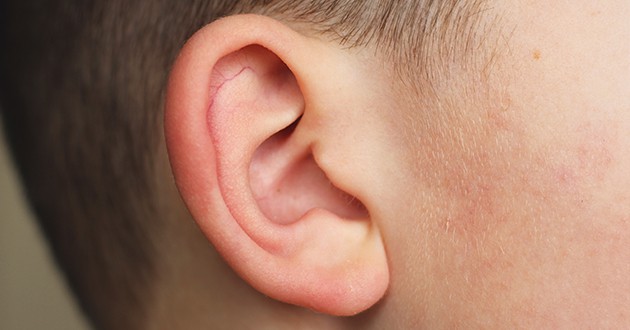
One in about 7,000 children in the world is born with a deformed earlobe (microtia) or with a complete lack of the earlobe (anotia). Microtia is more common in boys and is usually unilateral. If the other ear is normal, unhindered development of hearing and speech is enabled.
In children with microtia, an examination by an otorhinolaryngologist and a hearing test are necessary. Audiological processing includes tonal limited audiometry (at the age of 4 years), tympanometry and otoacoustic emission and BERA test.
Patients with unilateral microtia or atresia usually have normal hearing on the opposite side, and rehabilitation of these patients usually involves monitoring or possibly using hearing aids.
There are three ways to reconstruct the eardrum:
- Using prosthesis
- Using artificial material
- With the cartilage reconstruction of the patient
Ear prosthesis is an alternative to ear shell reconstruction. The operation is performed in one or two acts; the prosthesis is kept in position by osseointegration of titanium implants into the bone or by means of adhesive tapes. Children are bad candidates for prostheses, because constant visits to the doctor, maintenance of hygiene and frequent replacement of prostheses every two to five years are necessary. Prosthesis can be a good solution for adult patients, when the damage to the ear shell is a consequence of trauma or cancer.
Another possibility of ear shell reconstruction involves the use of artificial material and is possible from the third year of life. The advantage of this intervention is that there is no taking of costal cartilage, as well as achieving a good shape of the ear shell.
The best solution for the reconstruction of the auricle is autologous reconstruction, when the cartilage of the patient’s ribs is used to create the auricle. The possibility of tissue rejection of the patient is minimal.
The operation is performed in two acts:
- In the first act, which lasts between five and seven hours, the surgeon takes the rib cartilage, shapes the frame (ear) from it and places it under the skin. At the end of the first surgical procedure, two drains are placed in the operative wound, which extract the contents that collect in the vicinity of the prosthesis. The wound on the chest, where the cartilage is taken, leaves a minimal scar. Drains stand for up to four days. The patient received antibiotic and analgesic therapy. The length of stay in the hospital lasts up to three days. Thread removal after 15 days.
- Separation of the cartilaginous frame (second act) is performed at least six months after the first intervention. The procedure lasts up to three hours, and after lifting the cartilaginous frame, the area behind the ear is covered with skin flaps behind the ear shell and an autograft of the patient’s skin taken from the head or groin.
Interventions are performed by Dr. Aleksandar Urošević and Dr. Aleksandar Vlahović