


The esophagus is a tubular organ with the role of conducting chewed food and liquids from the oral cavity to the stomach. It consists of three parts and passes through the posterior part of the chest, behind the trachea, and in front of the aorta and the spinal column.
Considering its function, it is clear that esophageal disorders most commonly manifest as swallowing problems (dysphagia). The esophagus has a pronounced muscular layer that, at its beginning in the neck and transitioning into the stomach (in the abdomen), merges and forms a sphincter. This sphincter, in conjunction with the diaphragm, prevents the passage of acidic stomach contents back into the esophagus.
Inadequate function and relaxation of the lower esophageal sphincter can lead to food retention, as seen in patients with achalasia. In this article, we will further explain Esophagus illnesses as well as the procedures patients undergo for treatment.
Esophageal Cancer
The two most common esophagus cancers are squamous cell carcinoma and adenocarcinoma. They manifest similarly, but the risk factors are different. Basic risk factors for the formation of squamous cell carcinoma are alcohol consumption and smoking, excessive amounts of sour and spicy food, as well as being infected with the human papillomavirus (HPV).
Patients with achalasia have a higher long-term risk of developing squamous cell carcinoma, as do patients who have previously had laryngeal or oropharyngeal cancer. The main risk factors for adenocarcinoma are obesity and untreated reflux disease over multiple decades.
Symptoms of Esophageal Cancer
Patients often report feeling like food doesn’t pass through to the stomach or feeling like it stops in the chest. One of the other symptoms is intense unexplained weight loss over several months. Rarely, patients feel chest pain, commonly with bites of food that don’t go through the stomach. Every occasion of feeling like food stops in the chest and doesn’t go through to the stomach is a reason to visit a surgeon!
Diagnosis
During the diagnostic process, it is of utmost importance, if there’s the slightest doubt, to do an upper flexible endoscopy (gastroscopy), in order to adequately examine the mucosa of the esophagus, stomach, and duodenum.
In case of any type of abnormalities, biopsies are taken and then pathohistologically analyzed.
If esophageal cancer is confirmed, a CT scan of the neck, chest, and stomach should be done, with a double contrast (Oral and IV contrast). If there are doubts that the disease might be spreading, the doctor can suggest a stomach MRI, to further characterize the changes in the liver, or other methodical diagnostics (e.g., PET/CT scan).
If adenocarcinoma is confirmed before treatment initiation, the laboratory values of tumor markers (CEA, Ca 19-9, Ca 72-4) will be checked.
After completing the required diagnostics, patients are referred to an esophageal surgeon to determine the optimal treatment strategy. For locally advanced cases, individuals may be equipped with a nutritional stoma or undergo the placement of an esophageal stent for feeding. Subsequently, they are referred for neoadjuvant therapy aimed at reducing the stage of the disease (down-staging). If the disease is deemed resectable, surgery is then planned for the patients.
Hiatal Hernia
Hiatal hernia, or gastric hernia, is a very common condition where the stomach passes through the diaphragm into the chest. The frequency of this condition increases with age, and it is estimated that over 70% of patients over 70 years old have this condition.
Symptoms of Hiatal Hernia
There are sliding and paraesophageal hernias.
In sliding gastric hernias, patients usually report symptoms such as varying degrees of heartburn. In contrast, large mixed (paraesophageal) hernias, due to their size, lead to mechanical problems such as shortness of breath (due to occupying space in the chest), palpitations, and fatigue. In incarcerated hernias, nausea, vomiting, and food blockage after meals can occur.
Diagnosis
Patients with gastric hernia should first be referred for contrast radiography with barium. During the examination, the patient drinks contrast, and on the X-ray, the passage of contrast from the esophagus to the stomach can be observed, thereby showing the position of these organs in relation to the diaphragm.
It is also necessary to perform gastroscopy (upper flexible endoscopy) in order to assess the mucous membrane of the esophagus and stomach, exclude other possible conditions, and find objective indicators of the presence of gastric reflux.
In cases of suspected reflux disease, diagnosis should be supplemented with pH monitoring. This examination allows for the precise measurement of the time during which gastric contents are present in the esophagus.
A special group of patients complains of so-called extraesophageal symptoms, such as hoarseness, persistent cough, throat discomfort, and a feeling of a lump in the throat. A detailed conversation with the completion of precise evaluation questionnaires is necessary with this group of patients. It is important to identify with great care the groups of patients that respond best to therapy. By assessing the response to medication therapy, it is possible to evaluate which patients may benefit the most from the performed surgery.
Indications for surgical treatment include:
- Patients who are not motivated for lifelong intake of proton pump inhibitor medications.
- Patients with confirmed Barrett’s esophagus or severe esophagitis.
- Patients with persistent and pronounced symptoms despite the use of optimal medical therapy.
Achalasia and Esophageal Motor Disorders
Achalasia is a rare condition that occurs due to the inability of the lower esophageal sphincter to relax adequately. This condition leads to the retention of fluids and solid food in the esophagus, along with the eventual weakening of esophageal muscle activity during the progression of the disease.
The cause of the disease is not known, although it is believed to involve the deterioration of nerve centers within the esophageal wall that play a role in regulating muscle activity.
Symptoms of Achlasia
Patients with achalasia typically complain of food retention and weight loss. Food retention (dysphagia) usually progresses over several years, accompanied by a gradual, unwanted loss of body weight.
Diagnosis
When suspecting achalasia, it is crucial to use diagnostic methods to rule out malignant tumors of the esophagus and proximal stomach.
The diagnostic protocol typically begins with gastroscopy, during which we carefully examine the esophagus and its transition into the stomach. A common finding in patients with achalasia is the spasm (contraction) of the lower esophageal sphincter, making it challenging to pass the endoscope.
Contrast radiography with barium allows us to observe the dynamics of contrast medium passage through the esophagus, the time it stays in the lower part of the esophagus, as well as its diameter (width). It is also possible to observe the dynamics of the contraction of the muscular layer of the esophagus.
We complement the diagnostic process with manometry, which provides precise information about the condition of the muscular layer of the esophagus, its body, and the lower sphincter. Properly performed manometry allows for the classification of the type of achalasia.
Treatment of Achlasia
Temporary relief of symptoms in patients with achalasia can be achieved through pneumatic dilation, but a definitive solution can only be achieved through surgical treatment.
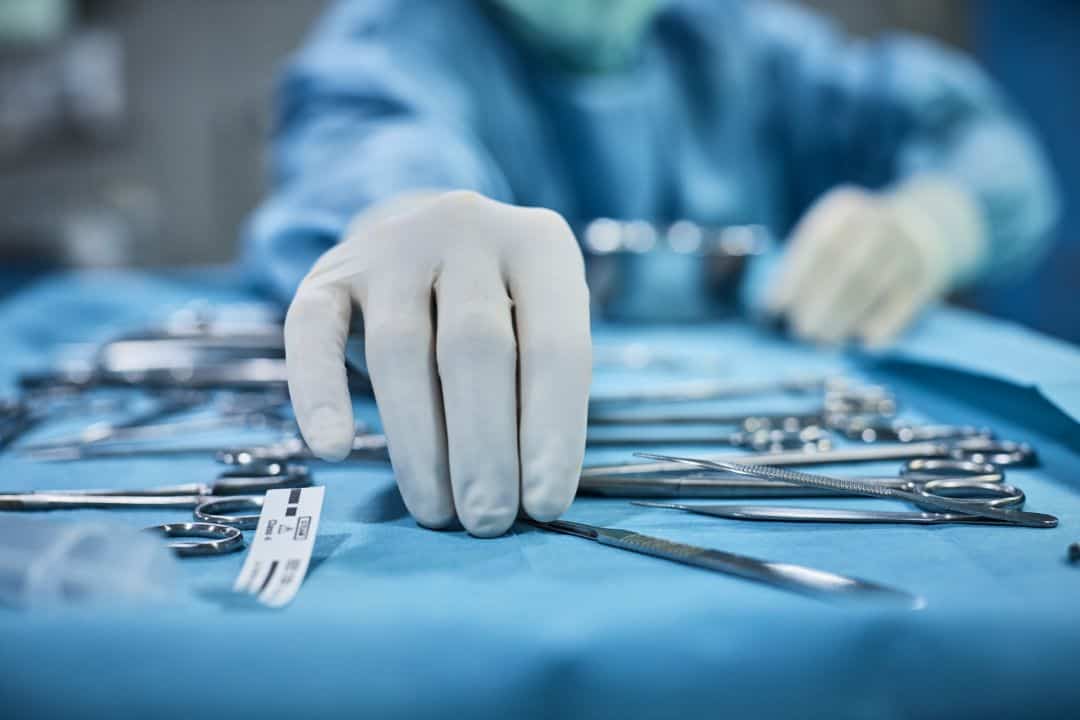
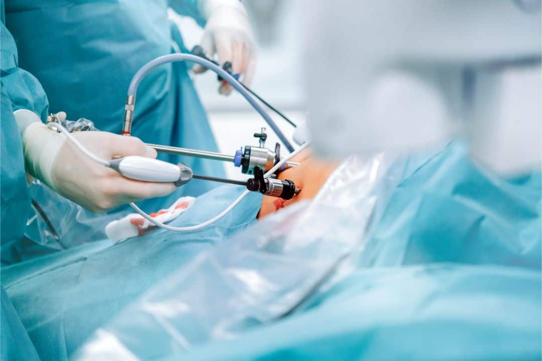
Minimal invasive surgery represents the gold standard in the treatment of the above-mentioned esophageal conditions.
Laparoscopy is a technique that provides access to the abdominal cavity through small incisions in the abdominal wall, typically ranging from 5 to 12 mm in size. Through specially created working channels (ports) and with the aid of sophisticated instruments and a camera with magnification of 5 to 10 times, we can access every part of the abdominal cavities and chest, effectively identifying and resolving the issue. These small incisions, compared to traditional surgeries, offer the potential for a rapid recovery, reduce the risk of infection, and allow a return to regular activities within a few days. The advantage of the magnified camera minimizes the possibility of errors in experienced hands, leading to the observation of the tiniest details during the procedure.
Thoracoscopy is a related procedure that allows us to access the thoracic cavity in the same way, through small incisions, without causing injury to the ribs and sternum.